Canal Foramen, Fissure, Space & Membrane: Topographical Anatomy and Image Diagnosis for Interpretation


Popis
Focusing on anatomical structures such as canals, foramina, fissures, spaces, and membranes, which have received less attention than solid organs, the book thoroughly explains where these structures are located, what they mean, and their significance in diagnostic imaging. Detailed explanations are provided with comprehensive diagrams and images to help you understand how experts in each field interpret these structures. This is an essential book for further improving your image interpretation and diagnostic skills.
[Major Topics] (For a detailed table of contents, click "View Table of Contents" below.) Chapter 1: Head and Neck
Overview—Skull Base Anatomy
Foramen Cecum
Cribrolamella Foramen
Optic Canal
Superior Orbital Fissure
Inferior Orbital Fissure
Pterygoid Fossa
Foramen Rotundum
Foramen Ovale
Carotid Canal
Foramen Lavatore
Facial Nerve Canal
Internal Auditory Meatus
Inferior Tympanic Canal
Foramen Magnum
Jugular Foramen
Hypoglossal Canal
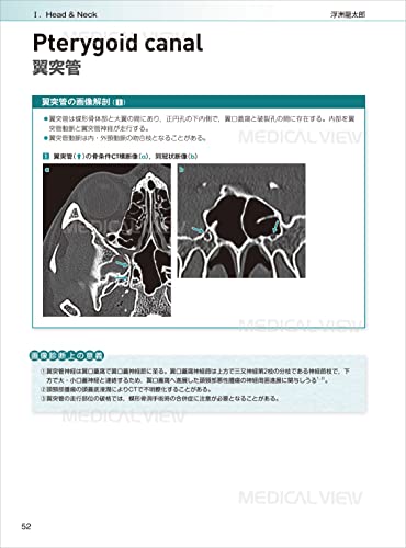
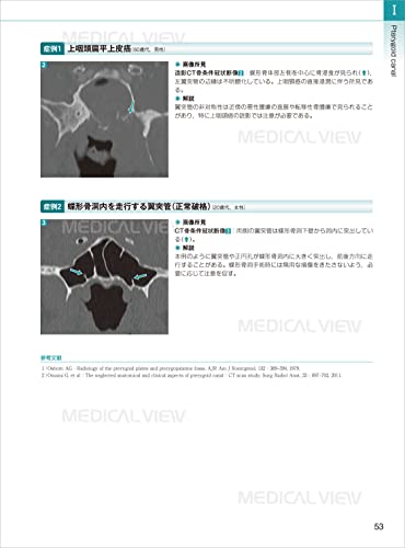
Pterygoid Canal
Foramen Spinus
Foramen Vesalius
Condylar Canal
Overview—Head and Neck Anatomy
Pharyngobasin Fascia
Parapharyngeal Space
Tensor-vascular Styloid fascia (TVSF)
Parotid space
Infratemporal fossa
Masseuse space
Cheek space
Sublingual space
Submandibular space
Carotid space
Retropharyngeal space
Danger space
Perivenx space
Pharyngeal mucosal space
Visceral space
Peritonsillar space
Posterior cervical space
Anterior epiglottic space, paraglottic space Chapter 2 Chest
Overview—Chest Anatomy
Anterior junctional line
Posterior junctional line
Azygos-esophageal recess
Left border of the descending thoracic aorta
Left paravertebral line
Right paratracheal line
Anterior aortic recess
Aortopulmonary window
Retrosternal space
Retrocardiac space
Line of Carly
Major interlobar fissure
Interlobular fissure
Accessory interlobular fissures: superior accessory interlobular fissure, inferior accessory interlobular fissure, left interlobular fissure, azygos fissure
Pulmonary ligament (mesopulmonary ligament)
Overview—Pericardial Anatomy
Transverse pericardial sinus
Oblique pericardial sinus
Superior pericardial cavity and left lateral recess Chapter 3 Abdomen and Pelvis
Overview: Anatomy of the Peritoneal Cavity
Subphrenic Space
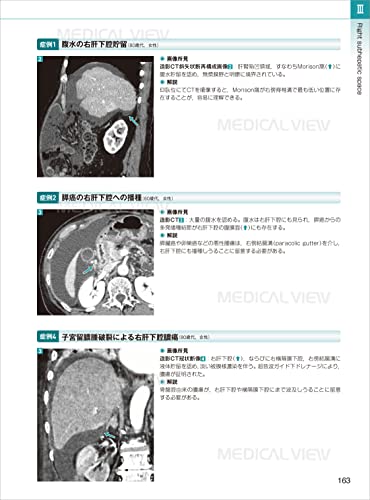
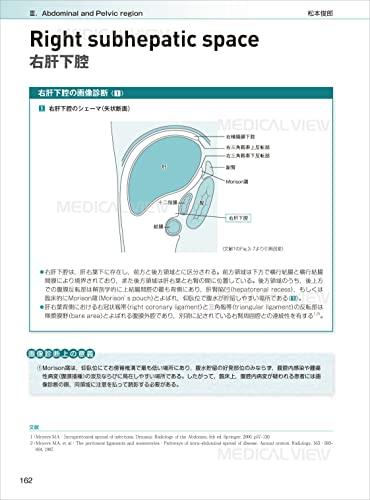
Right Subhepatic Space
Paracolic Groove
Omental Bag
Ligamenta Teres Hepatis
Hepatoduodenal Ligament
Hepatogastric Ligament
Gastrocolic Ligament
Gastrosplenic Ligament
Ligament of Treitz
Transverse Mesocolon
Sigmoid Mesocolon
Small Mesentery
Overview: Anatomy of the Retroperitoneal Space
Prerenal Space
Perirenal Space
Postrenal Space
Overview: Anatomy of the Pelvic Extraperitoneal Space
Prevesical Space
Perivesical Space
Perirectal Space
Presacral Space
Inguinal Canal
Femoral Canal
Obturator Foramen
Greater Sciatic Foramen Chapter 4 Bones, Joints, and Soft Tissues Overview - Shoulder Anatomy Acromion-to-head Distance Rotor Cuff Spacing Quadrilateral Space, Lateral Quadrangular Space Suprascapular Notch, Infraspinous Notch Shoulder Cavity (Including the Superior, Middle, and Inferior Glenohumeral Ligaments) Overview - Elbow Anatomy Cubic Tunnel Radial Nerve Canal Capitellar Fat Pad, Trochlear Fat Pad Synovial Plica (Elbow) Overview - Wrist Anatomy Carpal Tunnel Guyon Canal Extensor Finger Compartment Overview - Hip Anatomy Lesser Sciatic Notch Obturator Groove Piriform Fossa Ischiofemoral Distance Overview - Knee Anatomy Hoffa fat pad, infrapatellar fat pad
Synovial fold (knee)
Compartments separated by the medial collateral ligament
Meniscofemoral ligament, meniscotibial ligament
Pes anserine
Iliotibial band
Overview - Ankle Anatomy
Tarsal tunnel
Anterior tarsal tunnel
Sinus tarsi
Kager fat pad